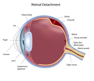
Retinal Tears and Detachment
Retinal Tears and Detachment: Information you need to know.
What is a retinal tear or hole?
Retinal tears occur when the retina begins to detach from the eye but is not yet completely detached.
Symptoms of a retinal tear or hole
Symptoms of retinal tears or holes may include flashes of light and eye floaters, where perceived specks occur in the field of vision.
Treatment for a retinal tear or hole
When a retinal tear or hole hasn’t yet progressed to detachment, our retinal disease specialist may suggest an outpatient procedure, which can usually prevent retinal detachment and preserve almost all of your vision.
Retinal tear treatment options can include:
- Laser Surgery
With laser surgery, the retinal disease specialist directs a laser beam at the retinal tear. The laser makes burns around the tear, creating scarring that usually “welds” the retina to underlying tissue. - Cryopexy
With cryopexy, the retinal disease specialist applies a freezing probe to the outer surface of the eye directly over the retinal defect and freezes the area around the hole, resulting in a scar that helps secure the retina to the eye wall.
Retinal Detachment
What is retinal detachment?
Retinal detachment is when the retina falls away or detaches from the rest of the eye.
What are the symptoms of retinal detachment?
Symptoms of retinal detachment may include bright flashes of light, hazy vision, and loss of vision in one eye.
Treatment for retinal detachment
If your retina has detached, the retinal disease specialist may use surgical procedures to repair it. Surgery doesn’t always work to reattach the retina. How well you see after surgery depends in part on whether the central part of the retina (macula) was affected by the detachment before surgery and, if it was, for how long. Your vision may take many months to improve after repair of a retinal detachment. Some people don’t recover any lost vision.
The specifics of your retinal detachment will determine which approach your retinal disease specialist recommends for treatment.
Treatment options for retinal detachment can include:
- Pneumatic Retinopexy—A surgical retinal disease specialist injects a bubble of air or gas into the vitreous space inside the eye. When the bubble is successfully placed to float against the retinal tear and the area surrounding the tear, it seals the tear. This stops further flow of fluid into the space behind the retina. Fluid that had collected under the retina is absorbed by itself, and the retina can then reattach itself to the back wall of your eye. You may need to hold your head in a certain position for up to several days to keep the bubble in place. The bubble eventually will also be reabsorbed on its own.
- Scleral Buckling—A surgical retinal disease specialist sutures a piece of silicone rubber or sponge to the white of your eye (sclera) over the affected area. The silicone material indents the wall of the eye and relieves some of the force caused by the vitreous tugging on the retina.
- Vitretomy—The surgical retinal disease specialist removes the vitreous gel along with any tissue that is tugging on the retina. Air, gas, or liquids are then injected into the vitreous space to reattach the retina.
FIVE CONVENIENT LOCATIONS
Ophthalmic Consultants of the Capital Region has five convenient locations to serve all your eye care needs. No need to travel to the big city. You can find advanced eye care right here in your hometown.
We have offices in:
Next Steps
If you’re experiencing blurred vision, floaters, or flashes of light, it is VERY important to call us immediately to schedule an appointment. Receiving prompt treatment can be critical to your sight.
Get in touch with us today at our nearest location to you.


