Overcoming Retinal Diseases: Can the Retina Heal Itself?

Dr. David Rabady
There are several retinal diseases that all lead to a varying degree of vision loss. Approximately 12 million Americans aged 40 and older suffer from vision impairment. But is retinal repair a possibility? Before we dive into that, let’s first deal with the different types of retinal damage.
Types of Retinal Conditions
There are several types of retinal damage such as:
- Retinal Tears and Detachment: These are the most common types of retinal damage as the retina pulls away from the tissue around it, leading to vision loss.
- Macular Holes: This is a tiny hole that develops in the macula (the central portion of the retina). These holes may be caused by multiple reasons, including eye trauma.
- Age-Related Macular Degeneration (AMD): AMD is a common eye condition in people aged 50 and above. It leads to a steady breakdown of the macula and loss of central vision.
- Vitreous Detachment: As people age, the vitreous gel in the eye shrinks. During this process, the vitreous gel may pull at the retina, leading to tears and possible detachment.
The Difference Between a Retinal Tear and Retinal Detachment
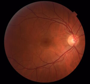
The retina is a light-sensitive layer of tissue that lines the eye’s interior and sends visual messages via the optic nerve to the brain. The larger back chamber of the eye is filled with a clear gel called the vitreous.
As stated before, a normal part of aging may lead to the vitreous gel shrinking and separating from the retina. You can develop a retinal tear as this gel pulls on the fragile retina and the process may also lead to retinal detachment.
Retinal tears may lead to bleeding inside the eye, blurring vision, and creating floaters. However, retinal tears can worsen into retinal detachment and vision loss.
How Does Retinal Detachment Occur?
The retina receives its oxygen and nutrition from a collection of blood vessels between itself and the wall of the eye. Once there’s a retinal tear or hole, the eye’s fluid moves through this opening and collects under the retina, detaching it from the eye’s wall. A detached retina is malnourished and unhealthy and eventually results in vision loss that could be permanent.
Causes of Retinal Tears and Detachment
Many people are interested in knowing what causes retinal tears and detachment. Almost all retinal detachments arise from vitreous separation due to normal aging.
Retinal tears may result from trauma and other diseases (although rare). If you have high myopia (extreme nearsightedness), then you are at an increased risk of a tear or detachment. Some individuals have weak areas in their retinas that may also increase their risk of having vitreous separation and retinal tears. In some cases, an eye doctor can treat specific areas to decrease the risks.
Kindly note that if you have a torn or detached retina in one eye, it heightens the risk of the same condition developing in the other eye. In almost all cases of retinal tears or detachment, the patient didn’t do anything to cause these conditions.
Symptoms of Retinal Tears and Detachment
Here are a few symptoms that will help you determine if you need to schedule an appointment with an eye doctor:
- Light flashes in one or both eyes
- The sudden appearance of new floaters or cobwebs
- Blurred vision
- A curtain-like shadow in your vision
If you experience any of these symptoms, please contact your eye doctor IMMEDIATELY.
The doctor will perform a dilated eye examination to closely inspect the retina. Retinal tears and detachments are often painless and so any corrective action has a much better vision outcome if performed before central vision is affected. A quick response to the warning signs may help prevent permanent vision loss.
Can a Damaged Retina Be Repaired?
Vision begins in the retina, the section of the eye that translates light into electrical signals to the brain. But when retina cells get damaged, they do not regenerate. They don’t heal or grow back.
The good news is that an eye doctor can repair a damaged retina in most cases. Although a patient may not experience completely restored vision, a timely retinal repair can prevent any further vision loss and stabilize vision.
However, it’s important to get treatment for a damaged retina as soon as possible. The earlier a patient accesses professional treatment, the better it is for their eyesight.
Top Retinal Repair Options
The good news is that there are several options to address retinal tears and detachment:
- Laser Photocoagulation: Laser procedure can often reseal a tear or reattach a torn section of the retina.
- Cryopexy: This is another method to reattach a torn segment of the retina. While laser photocoagulation uses thermal energy, cryopexy freezes the retinal tissue into place.
- Vitrectomy: This technique replaces the vitreous gel with a new substance to prevent further retinal damage. This procedure can be combined with any of the previous retinal treatment options.
- Scleral Buckling: This process involves sewing a small piece of silicone against the white of the eye. By creating an indentation on the eye’s surface, it relieves some of the tension of the vitreous gel pulling on the retinal tissue.
- Pneumatic Retinopexy: This procedure injects an air or gas bubble into the eye to help press the damaged retinal tissue into the right position to heal and reattach.
You don’t have to be alone in deciding on the best treatment option. Your eye doctor will closely examine your eyes to determine the most appropriate treatment process.
Exploring the Retinal Treatment Options

Once you suspect a torn or detached retinal diagnosis, you will need a specialist doctor right away! They will perform a retinal exam that includes using a bright light and special equipment and lenses to examine the back of the eye.
This specialized device offers a highly detailed view of the entire eye. It also helps them to closely examine your retina for any holes, tears, or detachment. Sometimes they may take photographs to help them fully document your retinal issues and make a well-informed diagnosis.
Treat Retinal Tears to Prevent Retinal Detachment
It is best to treat retinal tears before they worsen to become retinal detachment. Laser photocoagulation is often done in an ophthalmologist’s office. The specialist will use a special laser to surround the tear and seal it to prevent detachment.
In laser photocoagulation, a special lens focuses the laser (a concentrated light) on the retina to create small welds. Over time, these laser spots heal to hold the retina in place. After this short, in-office treatment, you will feel as though you were exposed to bright light directly into the eyes. However, the feeling returns to normal after a few minutes to hours. You will be happy to know that laser treatment can be done as often as needed.
Less frequently, the specialist uses cryopexy to freeze retinal tears and this procedure can be done in the doctor’s office. They will first use a numbing treatment to make the eye comfortable. Then they direct a freezing probe under the tear and deliver the freezing treatment around the tear. After treatment, the eye forms an adhesion that holds the retina in place.
Recovering From Laser Photocoagulation or Cryopexy
Many people recover well after retinal disease treatment. Patients can go home to relax in comfort, which aids the recovery process. Most patients will report that their eyes feel comfortable but dazed as though they had stared at an extremely bright light. However, these symptoms (including dilation) should wear off over the remainder of the day. Most people can go about their daily business post-operation and your doctor can advise you of the kinds of activities you can return to after the laser or cryopexy treatment.
In some rare instances, retinal detachment happens despite excellent laser treatment. This issue can arise if there are new tears and strong traction develops on the retina which makes the condition worsen.
Your retina specialist will carefully monitor your eye post-treatment to ensure you respond as expected to the retinal repair.
Managing Retinal Detachment Repairs
Timing is crucial for retinal detachment repair. Your retina specialist will attempt to fix the detachment before it impacts your central vision (or macula). If the macula is not yet affected, it will be vital to try and fix the retina urgently.
If the macula is impacted, the repair should occur within a few weeks. There are several repair methods and the option your doctor selects will depend on your specific case. Your doctor may even use a combination of treatments to attempt retinal restoration to improve vision.
Can Retinal Detachment Heal on Its Own?
It is rare for retinal detachment to go unnoticed by the patient and heal on its own. The vast majority of retinal detachments will progress to irreversible vision loss if left untreated. Therefore, it’s crucial to monitor any vision changes.
Vitrectomy Surgery for Retinal Repairs
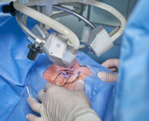
The eye surgeon will perform a vitrectomy in a special operating room with the patient under sedation. On the day of the surgery, they will ensure you are comfortable. The retina surgeon will remove the vitreous gel using small instruments via tiny holes in the sclera (the white part of the eye).
Once the retina is flattened, the surgeon applies the laser. Next, they place a gas bubble in the eye’s large chamber to temporarily hold the retina in place as the eye heals. There are two different types of gasses that can be used, lasting anywhere from two weeks to three months. In some cases, especially if the retina has formed scar tissue, they insert silicone oil into the eye to hold the retina in place. In most instances, no stitches are necessary. They then patch the eye with a bandage providing instructions to go home and relax.
Recovering From Vitrectomy Surgery
After a vitrectomy, you will need to hold your head in a certain position (it all depends on the locations of the retinal tears). You will have to restrict your activity level post-surgery, but there is little to no discomfort afterward. You will also need to take some eye drops to prevent infection and promote healing.
During the healing process, the gas bubble shrinks and your eye replaces it with its natural fluid. Also, your vision will improve gradually. While the gas bubble is inside your eye, it’s vital to stay off your back, not fly in an airplane, change altitudes, and inform your doctors if you need any other type of surgery.
Scleral Buckle Surgery for Retinal Detachment
This surgery is another option to correct retinal detachment. The procedure is done in a special operating room under general sedation. The eye surgeon will locate and treat the retinal damage using either laser photocoagulation or cryopexy. They then place a scleral buckle (or a special silicone band) around the eye and under the skin and muscles. The band gently pushes on the wall of the eye to keep the retina in place. Once the fluid drains from underneath the retina, they close the skin of the eye with dissolvable stitches.
In some cases, your doctor may inject a gas bubble to hold the retina in place from within the eye. Afterward, they will patch your eye with a bandage and let you go home to recover. Once again, you will need to hold your head in a certain position post-surgery (especially if a gas bubble was also inserted). Over-the-counter medications can help control any discomfort after surgery. You will also take prescription eye drops to prevent infection and promote healing.
Pneumatic Retinopexy for Retinal Detachment Repair
Pneumatic retinopexy is an in-office procedure used to correct retinal detachment. After the surgeon numbs the eye, they treat the retinal damage using laser photocoagulation or cryopexy and inject a gas bubble. The gas bubble floats and pushes the retina back into place to heal.
After the procedure, you are sent home to recover. As with the previous surgeries mentioned, you will need to keep your head in a certain position to hold your retina in place for optimal healing. You may also take prescribed eye drops to prevent infection and promote healing.
The Recovery Process After a Retinal Repair Surgery
Several aspects of retinal surgery recovery are determined by the type of surgery and the specific recovery time. Your doctor will give you recovery instructions specific to your case.
A big factor in your recovery time is the use of the gas bubble and the positioning required to keep the retina in place as you heal. There are two types of gas used in these bubbles — one lasts two weeks and another lasts two months. Your surgeon will choose the right gas bubble for your surgery and often try to use a faster-dissolving bubble. If silicone oil is placed in the eye, it is often removed several months after your eye has healed.
The majority of retinal detachments can be fixed with a single procedure. However, there are some cases where the retina gets detached again – usually from scar tissue developing. If the retina detaches once more, additional surgery is necessary.
Your vision may be blurry after the procedure but this condition gets better as the eye heals. Your doctor will monitor your condition very closely as your retina heals. But you also need to monitor your vision closely and immediately report any changes to your doctor.
Your retina doctor will also advise you about how much physical activity you can pursue immediately after the procedure. As well, be aware of a possible significant increase in floaters, flashes, or loss of peripheral vision.
Vision Outcomes After Retinal Detachment Repair
The outcomes may not be predictable after surgery and may not be known for several months afterward. Even after a successful procedure, your vision may not return to normal. The most significant factor in having good vision post-surgery is whether the central vision was damaged before the surgery.
Vision outcomes are best if you caught and addressed the retinal detachment before the macula (or center section of the retina) detaches. When the retina detaches, it separates from essential nutrients and oxygen supply, causing permanent damage to the retina.
Although surgery can reattach the retina, it can’t reverse the damage done to the retina. It is common to experience blurred or distorted vision which can’t be corrected with glasses, contacts, medications, or other types of surgery.
Frequently Asked Questions
Here are some frequently asked questions about retinal repair surgery:
Is retina damage permanent?
If you don’t treat retinal damage properly and immediately, permanent vision loss may occur. If you experience any symptoms of retina damage, contact Ophthalmic Consultants of the Capital Region right away.
How can I improve my retinal health?
You can improve the health of your retina by:
- Getting regular eye exams
- Quitting smoking
- Eating a healthy and balanced diet
- Wearing sunglasses when in sunny locations
- Wearing eye protection (especially when doing tasks)
- Drinking a lot of water
- Getting regular exercise
Can you drive after retinal detachment surgery?
Please consult with your retinal surgeon.
How do you sleep after a retinal detachment surgery?
If a gas bubble is used to fix your retinal detachment, staying off your back while sleeping is crucial. Sleeping on your stomach and sides is often allowed. However, your retina surgeon will advise you about the best way to sleep during your recovery.
How long does it take for a damaged retina to heal?
When your vision improves, it will be gradual. Complete healing after retinal surgery will take six months. In most cases, the visual acuity at six months will be your final vision. There will be normal swelling of the eye after retina surgery, which will limit your vision.
Get Expert Help for Your Eye Health
Are you or a loved one suffering from retinal diseases? Then we can help! At Ophthalmic Consultants of the Capital Region, our team of eye care experts offers treatment for different kinds of retinal conditions that can harm your vision. Whether dealing with a retinal tear, retinal detachment, or degenerative diseases, our world-class experts can help prevent further vision loss and improve general vision quality.


