About Retinal Disorders & Diseases
The retina is a very important thin layer of tissue on the inside back wall of your eye, containing millions of light-sensitive cells and nerve cells that receive and organize visual information. Your retina sends this information to the brain through your optic nerve, enabling you to see.
A retinal eye disease or disorder is an eye problem that affects the retina. When the retina is compromised, it can cause severe vision problems.
Ophthalmic Consultants of the Capital Region Retina Specialists
Ophthalmic Consultants of the Capital Region (OCCR) Retinal Specialists have treated thousands of patients with all types of retinal diseases and disorders. Experienced OCCR retinal specialists have advanced diagnostic tools and offer the latest treatments to restore vision or to slow or stop the disease and preserve as much of your vision as possible.
Retinal Disorders and Disease Causes
Retinal diseases can be associated with aging, diabetes, or other diseases, trauma to the eye, or family history.
Symptoms of Retinal Disorders Diseases
Symptoms might include seeing floating specks or cobwebs, blurred vision, distorted vision, defects in the field of vision, lost vision, or other problems.
Diagnosing Retinal Diseases
To make a diagnosis, one of our retinal disease specialists will conduct a comprehensive eye exam and looks for abnormalities in the eye.
Depending on your situation, they may perform the following tests:
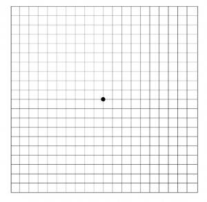
- Amsler grid test
Your retinal disease specialist at Ophthalmic Consultants of the Capital Region may use an Amsler grid to test the clarity of your central vision. They will ask you if the lines of the grid seem faded, broken, or distorted and will note where the distortion occurs on the grid to better understand the extent of your retinal damage. - Optical coherence tomography (OCT)
OCT is an excellent technique for capturing precise images of the retina to diagnose epiretinal membranes, macular holes, and macular swelling (edema), to monitor the extent of wet age-related macular degeneration and to monitor responses to treatment. Details of the cross-sectional anatomy of the retina can be displayed almost at a cellular level. - Fluorescein Angiography
Fluorescein angiography uses a dye that causes blood vessels in the retina to stand out under a special light. This helps to exactly identify leaking blood vessels, new abnormal blood vessels, subtle pigmentation changes in the back of the eye, signs of diabetic retinopathy, and other eye disorders. - Ultrasound
Ultrasound (ultrasonography) uses high-frequency sound waves to help view the retina and other structures in the eye, helping to distinguish cancerous (malignant) from noncancerous (benign) tumors. Also, an ultrasound can help “see-through” a dense cataract or a dense hemorrhage so that your retinal disease specialist can assess the retina and other structures.
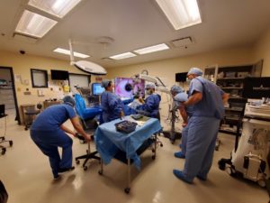
NGENUITY
Capital Region patients who may be suffering from a retinal condition or disease can choose Ophthalmic Consultants of the Capital Region with confidence for their eye care needs. In fact, the retinal group of surgeons at Ophthalmic Consultants of the Capital Region is the only retinal group to exclusively use the new NGENUITY 3D Visualization System with DATAFUSION technology during vitreoretinal surgery.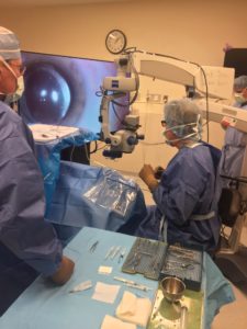
Vitreoretinal surgery is a sub-specialty of ophthalmology focused in diseases and surgery of the back of the eye including the retina, which focuses light and provides sharp, detailed vision, and the vitreous body of the eye, which is a clear gel that fills the space between the retina and the lens.
The NGENUITY system used by our surgeons is made up of a 3D stereoscopic, high-definition digital video camera and workstation that provides magnified stereoscopic images of objects during microsurgery. The system allows surgeons to perform “heads-up” surgery, operating while looking up at a high-definition 3D monitor instead of bending their necks to look through the eye-piece of a traditionally used operating microscope. This allows retinal surgeons unprecedented 3D visualization of the back of the eye with greater depth and detail during surgery than ever before.
The NGENUITY 3D Visualization System with DATAFUSION technology allows surgeons several improvements over traditional microscopes, including:
- a greater depth of field, allowing for a larger working space at the retina surface, in high magnifications;
- high-dynamic range imaging that allows the surgeon to reduce the glare of instruments, and also to see into the shadows;
- lower light levels of 50% or more reduce phototoxicity to both patient and surgeon;
- use of digital image filtering, which allows for different color effects, assisting surgeons in augmenting contrast.

The system also allows our retinal surgeons to track key elements in real-time during procedures, such as intraocular pressure, flow rates, infusion pressure, and laser power—meaning that retinal surgeries are safer and more effective than they’ve ever been before.
Our retinal surgeons care about your eyes and providing you with the best possible results, which is why Ophthalmic Consultants of the Capital Region continually adopts the best new advancements in eye care, including the full-time use of the NGENUITY system.
FIVE CONVENIENT LOCATIONS
Ophthalmic Consultants of the Capital Region has five convenient locations to serve all your eye care needs. No need to travel to the big city. You can find advanced eye care right here in your hometown.
We have offices in:
Next Steps
If you’re experiencing blurred vision, floaters, flashes of light, or have been diagnosed with diabetes, it is important to call us immediately to schedule an appointment. Receiving prompt treatment can be critical to your sight.
Get in touch with us today at our nearest location to you.