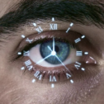
What is Glaucoma? Exploring the Symptoms, Diagnosis, and Treatments
What is glaucoma?
Glaucoma is a prevalent eye disease worldwide and is the second leading cause of vision loss. Approximately 3 million Americans have glaucoma, but half of these individuals may not know that they have the disease.
This lack of awareness may be due to glaucoma initially having no symptoms. In fact, early vision loss is often not recognized by patients. Glaucoma has been called the “silent thief of sight” because it typically causes no symptoms until the disease is very advanced. While this condition can’t be cured, you may take early action to preserve your vision and prevent vision loss. Let’s delve into the details of all things glaucoma:
What is Glaucoma?
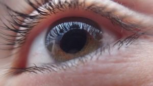
Glaucoma is an eye disease that causes chronic and progressive damage to the optic nerve. This optic nerve transmits visual information from your eyes to your brain. There are many factors that cause glaucoma, including high eye pressure, age, and genetics. Significant optic nerve damage results in permanent vision loss. There are currently no treatments to reverse vision loss from glaucoma. But the good news is that if you catch glaucoma early, you may be able to prevent additional vision loss.
Glaucoma Quick Facts
- Glaucoma is the leading cause of irreversible blindness worldwide.
- Although anyone can develop glaucoma, certain individuals are at greater risk.
- Early diagnosis and treatment are crucial in preserving sight in glaucoma patients.
- Glaucoma is an eye disease often linked with higher intraocular pressure (IOP). This elevated pressure damages the optic nerve in the eye, leading to vision loss and possibly blindness.
- IOP rises when the eye produces too much fluid or the eye’s drainage or outflow channels (trabecular meshwork) gets blocked.
- The two main types of glaucoma are:
- Open-angle glaucoma. There are several variants and is chronic (or long duration).
- Angle-closure glaucoma. It may be either acute (sudden) or a chronic condition.
- The condition often causes no symptoms at first. At that stage, glaucoma is only diagnosed by regular eye exams.
- Testing for glaucoma is painless and assesses the presence of the condition and its progression. These tests assess:
- Level of IOP
- Optic nerve status
- Drainage angle
- Visual fields
- Glaucoma damage to the optic nerve and vision impairment is irreversible.
- The best known treatment of glaucoma is lowering of the eye pressure. There are three ways to lower IOP: medication (eye drops), laser, and surgery. Most cases can be controlled quite well with these treatments, preventing further vision loss.
- There are many ongoing glaucoma clinical studies worldwide.
What Are the Glaucoma Symptoms?
The most common glaucoma type is primary open-angle glaucoma. It has no signs except for gradual vision loss. Other symptoms of late stage glaucoma include glare and difficulty adjusting to changes in room lighting. Therefore, you should get annual comprehensive eye exams so your ophthalmologist can monitor for changes in your vision.
Acute angle-closure glaucoma (or narrow-angle glaucoma) is a medical emergency. Contact a physician right away if you experience any of these glaucoma symptoms:
- Eye redness
- Severe eye pain
- Sudden blurred vision
- Seeing colored rings around lights
- Sudden vision disturbances
- Nausea
- Vomiting
What Causes Glaucoma?
The eye continuously makes aqueous humor, a clear fluid. As this fluid is produced, it circulates to the front of the eye. It then leaves your eye through the trabecular meshwork, which is located in the angle of the eye. The angle is between the cornea and iris. From the trabecular meshwork, the aqueous humor drains into outflow channels that empty into the veins of the eye. If these channels are partially or fully blocked, the natural pressure in your eye (IOP) may increase. As the IOP rises, your optic nerve may get damaged and progressively worsen, causing vision loss.
What Causes an Increase in Intraocular Pressure?
The following factors can cause the IOP to increase:
- Restricted or blocked drainage in the eye
- Steroid medications
- For patients at risk of angle closure glaucoma, medications or drops that cause dilation
What Are the Types of Glaucoma?
There are four major glaucoma categories:
Open-Angle (Chronic) Glaucoma
According to the National Eye Institute (NEI), open-angle glaucoma is the most common type of glaucoma. This type of glaucoma has few or no signs except gradual vision loss. This loss is often slow so that your vision suffers irreparable harm before any other symptoms become visible. Risk factors for open angle glaucoma include family history, elevated eye pressure, thin corneas, and race (higher in African Americans and Hispanics).
Angle-Closure (Acute) Glaucoma
This type of glaucoma is characterized by a narrow angle anatomic configuration. In other words, the shape of the eye is a risk factor for angle closure glaucoma. It is usually diagnosed in younger people but can get worse as patients get older. This is because cataracts cause the angle to get more narrow over time. In patients with narrow angles, the iris is abnormally close to the cornea. If the pupil dilates, the iris can block off and close the angle. Aqueous humor builds up in the eye because it cannot access the trabecular meshwork (drainage outflow).
Angle closure can happen acutely or slowly over time. If it happens suddenly, the rapid accumulation of aqueous humor often causes a painful increase in eye pressure. This acute angle-closure glaucoma is an emergency, and you should call your doctor right away if you start having symptoms like blurred vision, nausea, and severe pain. Risk factors for angle closure glaucoma include family history, far-sightedness, and race (higher in Asians, Hispanics and Caucasians).
Congenital Glaucoma
Children with congenital glaucoma are born with a defect in the outflow pathway of the aqueous humor: the angle and trabecular meshwork. Aqueous humor is unable to drain through the abnormal tissue. Parents will notice that their baby’s eyes will appear cloudy, or tear or blink to light. This type of glaucoma may also run in families.
Secondary Glaucoma
Secondary glaucoma is a side-effect of injury or another eye pathology like cataracts, inflammation, and eye tumors. Medicines (for example, corticosteroids) may also lead to glaucoma. Eye surgery may also cause secondary glaucoma.
Who is at Risk of Glaucoma?
Some of the major significant factors for glaucoma include:
Age
People above 60 years are at greater risk of glaucoma, which increases slightly each year. However, African-Americans face a greater risk of glaucoma beginning at 40 years old.
Race/Ethnicity
African-Americans (or people of African descent) are more likely to develop open angle glaucoma from high eye pressure than Caucasians. Caucasians may have pseudoexfoliation syndrome or pigment dispersion syndrome, which is a risk factor for glaucoma. Furthermore, Asian-descent people have a higher risk of angle-closure glaucoma.
Eye Issues
Chronic inflammation in the eye and thin corneas could lead to increased IOP. Physical injury or eye trauma (like being hit in the eye) may also lead to greater eye pressure.
Family History
Some glaucoma types are hereditary. Therefore, if a parent or grandparent had open-angle glaucoma, you are at a higher risk of developing the eye condition.
Medical History
People with diabetes, high blood pressure, and heart disease are at a greater risk of developing glaucoma. Furthermore, using certain medicines (such as corticosteroids) for extended periods could increase the risk of developing secondary glaucoma.
Glaucoma Diagnosis: The Evaluation Process
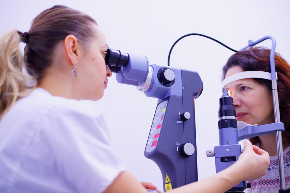
An ophthalmologist (an eye doctor) can often detect those persons at risk of glaucoma before nerve damage occurs. For example, the specialist may detect a narrow drainage angle or increased IOP.
The doctor may also diagnose patients with glaucoma by observing the extent of their nerve damage or visual field loss. The following six tests (all painless) may also be a part of this evaluation.
Tonometry
This test measures IOP by assessing the tone or firmness of its surface. Several tonometers exist for this test. However, the most common one is the applanation tonometer.
Once the ophthalmologist numbs the eye with anesthetic eye drops, they place the tonometer’s sensor against the eye’s front surface. The firmer the eye’s surface tone, the higher the pressure reading.
Pachymetry
What is pachymetry? It measures the thickness of the cornea (the front surface of the eye). Once the eye is numbed with anesthetic eye drops, the ophthalmologist lightly touches the pachymetry tip to the cornea.
Studies indicate that corneal thickness impacts the measurement of the IOP. Thicker corneas may give false high-pressure readings, while thinner corneas may give false low-pressure readings. Thinner corneas are a glaucoma risk factor. Once your doctor knows the thickness of your cornea, they can accurately interpret your tonometry.
Gonioscopy
In gonioscopy, the doctor numbs the eye with anesthetic drops and places a special contact lens with mirrors on the eye’s surface. What’s the purpose of these mirrors? They help the doctor view the eye’s interior from several directions.
This test examines the angle, which contains the trabecular meshwork. The trabecular meshwork is the tissue that drains the aqueous humor from the eye. The ophthalmologist determines whether the angle is open or narrow or if there are any other abnormalities. Some of these abnormalities include increased pigment within the angle or long-term damage to the angle arising from previous inflammation or injury.
People with narrow angles may have an increased risk for sudden onset of angle-closure, leading to an acute angle-closure glaucomatous attack. Gonioscopy may also determine if the eye is vulnerable to chronic angle closure, if the blood vessels are abnormal, and if hidden tumors are blocking aqueous fluid draining from the eye.
Ophthalmoscopy
Ophthalmoscopy is where the doctor uses a handheld device, a head-mounted device, or a special lens and slit lamp for the eye examination. Then, the doctor looks directly through the pupil (the opening in your colored iris) into your eye. The objective is to examine the optic nerve at the back of the eye for any damage.
An ophthalmoscopy exam detects any cupping of the disc (or damage to the optic nerve). Cupping refers to an indentation of the optic disc generally arising from increased intraocular pressure. Asymmetry in the extent of optic nerve cupping between both eyes may be an indicator of glaucoma. Furthermore, the optic nerve cupping may increase over time. Also, the pale color of the nerve may indicate damage due to poor blood flow or higher intraocular pressure. It’s possible to compare changes to the optic nerve over time as photographs can be taken of the optic nerves to facilitate comparison.
Visual Field Testing
This procedure maps the visual fields to detect any early or late signs of glaucoma damage to the optic nerve. Each visual field is analyzed by a computer one eye at a time to diagnose and monitor any glaucoma present.
The technician covers one eye, and the patient places his/her chin in a bowl. Next, lights of varying intensity are randomly projected within the bowl’s interior. Whenever the patient sees the light, he/she presses a button. Doing so creates a computerized map of that patient’s visual field. It also outlines areas where each eye can or cannot see. With glaucoma, there are characteristic changes in the visual field.
Confocal Laser Scanning Systems
Optical coherence tomography is a non-invasive imaging system. A machine with a camera creates a three-dimensional image of the optic nerve and retina to assess specific characteristics. These characteristics include the degree of cupping and the thickness of the retinal nerve fiber layer (RNFL). Thinning of the RNFL can indicate glaucoma. The results of OCT scans help assess and quantify any ocular damage arising from all types of glaucoma.
What Are the Glaucoma Treatment Options?
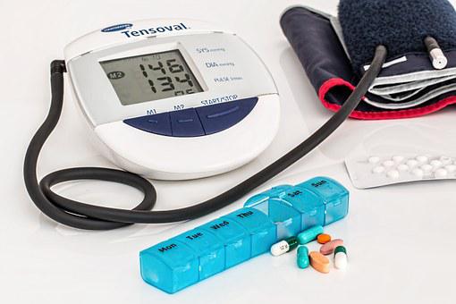
The objective of glaucoma treatment is to decrease IOP, which halts any further vision loss. Generally, your doctor will commence treatment using prescription eye drops. If these do not work, then they may recommend one of these treatments:
Medications
Several medications to reduce IOP are available in eye drops and pills. Your ophthalmologist will recommend the best one for your case.
Laser
There are two commonly performed procedures for treatment of glaucoma. Selective laser trabeculoplasty (SLT) applies laser energy to the trabecular meshwork. This allows opening of the tissue to increase the amount of drainage of aqueous humor. The increase in outflow results in lowering of the intraocular pressure.
Laser peripheral iridotomy is performed in patients with narrow angles who are at high risk or show signs of angle closure. Laser energy is used to create a small opening in the peripheral iris. This procedure helps prevent acute angle closure glaucoma.
Surgery
If medication and laser treatments are not enough to lower the IOP, surgery may be the best recommendation. The most commonly performed surgeries for glaucoma include trabeculectomy and tube shunt insertion.
Both surgeries create an outflow pathway that bypasses the eye’s dysfunctional tissue. Trabeculectomy surgically creates the outflow pathway through the cornea and sclera. Alternatively, a tube shunt is a device that can be implanted in the eye. The tube is placed in the eye to allow drainage of the aqueous humor. Both of these surgical options are incisional, meaning the eye tissue is cut and manipulated in the surgical field.
An alternative to incisional surgery is laser surgery, which is performed as transscleral cyclophotocoagulation. During this laser procedure, a probe is moved across the surface of the eye to ablate, or destroy, the ciliary body. The ciliary body is the tissue in the eye that produces the aqueous humor. Destruction of the ciliary body results in lowering of the IOP.
Does Glaucoma Cause Blindness?
Yes, glaucoma can cause blindness. However, complete blindness (loss of light perception in both eyes) is uncommon. It is possible to slow or stop vision loss from glaucoma with the above mentioned treatments and appropriate monitoring. However, there is no glaucoma cure, and you will likely need regulatory IOP treatment for life. Unfortunately, once there is vision loss, it is irreversible.
How Can We Prevent Glaucoma?
While we can’t prevent glaucoma, it is essential to have an early diagnosis to initiate appropriate treatment and monitoring, which will help slow progression and prevent vision loss that could happen over the course of your life. What’s the best way to manage this condition? Have an annual preventive eye care appointment with your ophthalmologist. Simple eye tests may detect glaucoma damage before it progresses and causes vision loss during these routine visits.
The Future of Glaucoma Studies
The frontier of glaucoma research is ever-evolving. Eye drops are available for glaucoma treatment. Some are innovative agents, while others combine new formulas with existing ones. The latter type of eyedrops is more affordable for patients. Currently, research is conducted at the National Eye Institute and most medical-school-affiliated institutions.
Lowering IOP is the primary way to treat glaucoma. However, some experts consider the disease as mainly a neurological condition. Therefore, researchers are investigating the neuroprotection of the optic nerve, especially in patients who appear to suffer from progressive nerve damage and visual field loss (despite relatively normal IOPs).
Researchers discovered that animal models showed that specific chemical mediators could decrease injury or death of nerve cells. However, biopsy or tissue specimens from humans are not easily available, so it is difficult to prove the benefit to the human optic nerve. Nevertheless, if any eye drop-based mediators can protect the human optic nerve from glaucomatous damage, they would significantly prevent blindness.
As well, new surgeries are being evaluated to see how they can safely lower IOP without the great possibility of damage to the eye or vision loss. There are also increased efforts to build public awareness of the impact of glaucoma, free screenings worldwide, early diagnosis and treatment, and treatment compliance.
These combined efforts are our best chance to minimize glaucoma-related vision loss.
Frequently Asked Questions (FAQs)

Here are some of our FAQs:
Is there a cure for glaucoma?
There is no cure for glaucoma, but the disease can be stabilized. Eyedrops, pills, laser treatments, and surgical procedures can prevent or slow down further damage. Regardless of glaucoma, regular eye exams are crucial to detect glaucoma progression and prevent vision loss.
What’s the glaucoma timeline?
Approximately 15 percent of glaucoma cases may lead to blindness in at least one eye over 20 years. Fortunately, glaucoma often progresses slowly over the years.
How do I know if I have glaucoma?
Vision loss, glare, difficulty adjusting to changes in light settings, such as going from a dark room to light could be signs of glaucoma. Again, these symptoms only occur once glaucoma is in its advanced, or late stage.
Some of the signs of acute angle-closure glaucoma are:
- Blurred vision
- Red eyes
- Halos around lights
- Severe eye pain
- Nausea and vomiting
- Sudden occurrence of visual disturbance (especially in low light)
What foods should I avoid if I have glaucoma?
Eating foods high in trans fatty acid can damage the optic nerve. Therefore, we recommend that you forego baked goods like cookies, cakes, donuts, fries, or stick margarine to avoid worsening your case and boost your eye health.
Does glaucoma ever go away on its own?
Glaucoma never gets better; it can either stabilize or get worse. Glaucoma is a chronic condition; therefore, any patient diagnosed with glaucoma needs lifelong monitoring for progression. It is possible for glaucoma to stabilize and not progress, but any nerve damage that has occurred does not improve.
Glaucoma is often far advanced when a patient becomes aware of vision loss. Such vision loss is not reversible even with advanced treatment such as surgery. Since open-angle glaucoma has few warning signs before damage has occurred, it’s crucial to visit a physician for regular eye examinations.
Choose the Vision Experts
We have explored the question “What is glaucoma?” and the symptoms, diagnosis, and treatment options. Ophthalmic Consultants is your best treatment source to address glaucoma and other eye conditions. Contact us today to discuss how we can help you maintain your vision.